By using fruit fly as a model system, Minna Poukkula working at the Institute of Biotechnology, University of Helsinki, has now elucidated how actin-rich protrusions contribute to cell migration in animal tissues. She revealed that GMF, a protein that promotes the disassembly of branched actin filament networks, controls the size and lifetime of protrusions in border cell clusters migrating in fruit fly egg chambers. Importantly, diminished protrusion dynamics in GMF-deficient flies correlated with problems in border cell cluster migration.
"These findings demonstrate that efficient actin filament disassembly by GMF is essential for rapid dynamics of cell protrusions, and that this dynamics are important for cell migration in a three-dimensional tissue environment", says Minna Poukkula from the research group of Pekka Lappalainen.
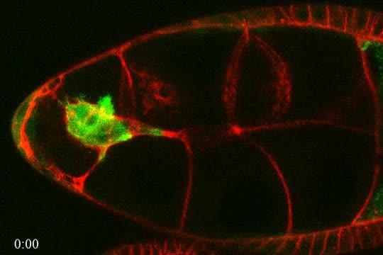
Fruit fly border cells form clusters of six to eight cells, which display directional migration during oogenesis. Migration of border cells in egg chambers can be examined in detail by live-cell microscopy. The egg chamber is in red and border cells (their actin cytoskeletons) are in green. The border cells display protrusions that are important for their migration in the tissue environment. The movie represents an approximately a 2-hour time window of a fruit fly egg chamber.
(Photo Credit: Pekka Lappalainen group, University of Helsinki)
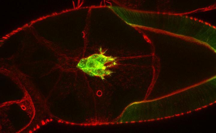
Fruit fly border cells form clusters of six to eight cells, which display directional migration during oogenesis. The egg chamber is in red and border cells (their actin cytoskeletons) are in green. The border cells display protrusions that are important for their migration in the tissue environment.
(Photo Credit: Pekka Lappalainen group, University of Helsinki)
Source: University of Helsinki