The scientists start with a so-called hydrogel – a material made of macromolecules, arranged in a loose meshwork. Between those molecules, large pores remain, through which other molecules or even cells can migrate.
Specially selected molecules are introduced into the hydrogel meshwork, then certain points are irradiated with a laser beam. At the positions where the focused laser beam is most intense, a photochemically labile bond is broken. That way, highly reactive intermediates are created which locally attach to the hydrogel very quickly. The precision depends on the laser's lens system, at the Vienna University of Technology a resolution of 4 µm could be obtained. "Much like an artist, placing colors at certain points of the canvas, we can place molecules in the hydrogel – but in three dimensions and with high precision", says Aleksandr Ovsianikov.
Chemical Signals for Cells
This method can be used to artificially grow biological tissue. Like a climbing plant clinging to a rack, cells need some scaffold at which they attach. In a natural tissue, the extracellular matrix does the trick by using specific amino acid sequences to signal the cells, where they are supposed to grow.
In the lab, scientists are trying to use similar chemical signals. In various experiments, cell attachment could be guided on two dimensional surfaces, but in order to grow larger tissues with a specific inner structure (such as capillaries), a truly three dimensional technique is required.
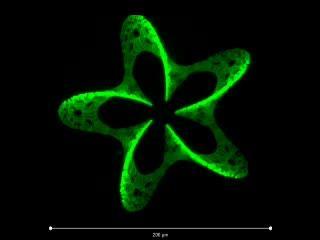
This is a 3-D pattern produced by photografting (180 µm wide). Fluorescent molecules are attached to the hydrogel, resulting in a microscopic 3-D pattern.
(Photo Credit: Vienna University of Technology)
Micro Sensors Detect Molecules
Depending on the application, different molecules can be used. 3D photografting is not only useful for bio-engineering but also for other fields, such as photovoltaics or sensor technology. In a very small space, molecules can be positioned which attach to specific chemical substances and allow their detection. A microscopic three-dimensional "lab on a chip" becomes possible.Weblinks
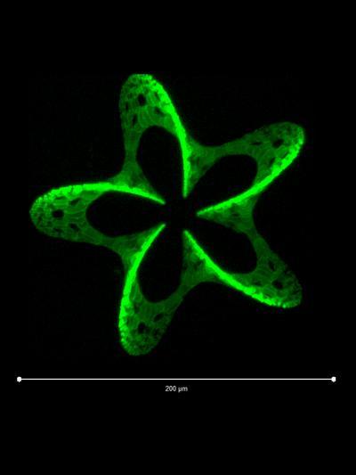
This is a 3-D pattern produced by photografting (180 µm wide). Fluorescent molecules are attached to the hydrogel, resulting in a microscopic 3-D pattern.
(Photo Credit: Vienna University of Technology)
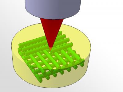
A laser shines into the hydrogel (yellow), attaching molecules to it at specific points in space (green).
(Photo Credit: Vienna University of Technology)
Source: Vienna University of Technology