When brain cells were injected into the surrounding gel, the cells released chemicals that prompted the engineered vessels to sprout new branches, extending the network. A similar system could supply blood to engineered tissue before transplant into the body.
After joining the UW last year, Zheng collaborated with the Puget Sound Blood Center to see how this research platform would work to transport real blood.
The engineered vessels could transport human blood smoothly, even around corners. And when treated with an inflammatory compound the vessels developed clots, similar to what real vessels do when they become inflamed.
The system also shows promise as a model for tumor progression. Cancer begins as a hard tumor but secretes chemicals that cause nearby vessels to bulge and then sprout. Eventually tumor cells use these blood vessels to penetrate the bloodstream and colonize new parts of the body.
When the researchers added to their system a signaling protein for vessel growth that's overabundant in cancer and other diseases, new blood vessels sprouted from the originals. These new vessels were leaky, just as they are in human cancers.
"With this system we can dissect out each component or we can put them together to look at a complex problem. That's a nice thing—we can isolate the biophysical, biochemical or cellular components. How do endothelial cells respond to blood flow or to different chemicals, how do the endothelial cells interact with their surroundings, and how do these interactions affect the vessels' barrier function? We have a lot of degrees of freedom," Zheng said.
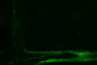
This video shows blood pumping through an engineered microvessel stimulated with an agent that causes inflammation. Over time green clots form in the vessel, like they do in the human body.
(Photo Credit: Y. Zheng, U. of Washington)
The system could also be used to study malaria, which becomes fatal when diseased blood cells stick to the vessel walls and block small openings, cutting off blood supply to the brain, placenta or other vital organs.
"I think this is a tremendous system for studying how blood clots form on vessels walls, how the vessel responds to shear stress and other mechanical and chemical factors, and for studying the many diseases that affect small blood vessels," said co-author Dr. José López, a professor of biochemistry and hematology at UW Medicine and chief scientific officer at the Puget Sound Blood Center.
Future work will use the system to further explore blood vessel interactions that involve inflammation and clotting. Zheng is also pursuing tissue engineering as a member of the UW's Center for Cardiovascular Biology and the Institute for Stem Cell and Regenerative Medicine.
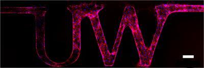
The engineered vessels can be made into any shape. In this case, the researchers made a model in the shape of the University of Washington logo. The white bar is 100 micrometers, about the width of a human hair.
(Photo Credit: Y. Zheng, U. of Washington)
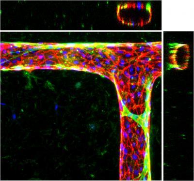
The engineered microvessels can form bends and T-junctions. The blue dots are the nuclei of the endothelial cells in the vessel walls, and the cell junctions are red. When smooth muscle cells (green) are introduced, they wrap and tighten around the vessels like they do in the human body.
(Photo Credit: Y. Zheng, U. of Washington)
Source: University of Washington