New simple and effective methods are needed to better evaluate the outcomes of repair using nerve conduits in vivo. Ultrasound is a common noninvasive clinical detection modality that has been used in many fields. However, ultrasound has only rarely been used to observe implanted nerve conduits in vivo. Hongkui Wang and co-workers from Affiliated Hospital of Nantong University report the first use of ultrasound to noninvasively observe the changes in chitosan nerve conduits implanted in rats over time. The ultrasound imaging clearly showed whether there are unsatisfactory complications after implantation, such as fracture, collapse, bleeding, or unusual swelling of the nerve conduits; and reflected the degradation mode of the nerve conduit in vivo over time. Ultrasound, as a noninvasive imaging modality, can be used as a supplementary observation method during conventional animal experiments on peripheral nerve tissue engineering. The relevant study has been published in the Neural Regeneration Research (Vol. 9, No. 14, 2014).
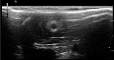
This is an ultrasound image of the morphology of a chitosan nerve conduit in a rat model of sciatic nerve defects at three weeks after modeling.
(Photo Credit: Neural Regeneration Research)
Source: Neural Regeneration Research