When the researchers eliminated both BRCA1 and p53, they found the neurons grew at a normal rate, but still disorderly, with cells pointed in the wrong direction.
"In this scenario, we recover a lot of neurons but there's still a lot of abnormalities, such as cells that are sideways and pointed the wrong direction," says Gerald Pao, who, along with Quan Zhu and Perez–Garcia, is a primary contributor to the paper and Salk researcher.
Scientists at the Salk Institute have uncovered details into a surprising -- and crucial -- link between brain development and BCRA1, a gene whose mutation is tied to breast and ovarian cancer. Video: Courtesy of the Salk Institute for Biological Studies
This observation led the team to propose that BRCA1 has an additional role in assisting neurons in orienting: the gene acts on the centromere of DNA—essentially an anchor for the chromosome arms essential in cell replication—to tell the new cell in which direction to grow, providing guidance in developing the brain's organized layers.
"It is remarkable that BRCA1 has such a significant effect on the brain, especially size. This work leads us to a better understanding of how to protect neurons," says Verma, who is also the Irwin and Joan Jacobs Chair in Exemplary Life Science. Because BRCA1 seems to regulate the centromere, studying the gene will help scientists to understand how mammalian brains have evolved over time.
"Now we have an explanation for why some patients with breast cancer also experienced brain seizures," adds Pao. This knowledge could potentially help identify breast cancer–susceptible patients predisposed to seizures and provide appropriate treatments.
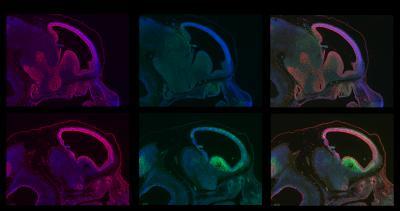
The images show an early developmental stage of normal (top row) and BRCA1-deficient brains (bottom row). The imaged embryos show abundant proliferation of cell growth (red, first column) in both normal and BRCA1-deficient brains at this stage. However brains lacking BRCA1 exhibit high levels of cellular suicide (green, second column). The third column shows an overlay of the other columns.
(Photo Credit: Image: Courtesy of the Salk Institute for Biological Studies)
Source: Salk Institute