CHAMPAIGN, Ill. — An ancient Egyptian mummy has had quite an afterlife, traveling more than 6,000 miles, spending six decades in private hands, and finally, in 1989, finding a home at the World Heritage Museum (now the Spurlock Museum) at the University of Illinois. The mummy's travels did not end there, however. It has made two trips to a local hospital – once in 1990 and again this year – for some not-so-routine medical exams.
Egyptologists, a radiologist, a pathologist, a physical anthropologist and a mummy expert are using the best diagnostic tools available to learn about the mummy without unwrapping its red linen shroud or cutting into it. The team will discuss its findings during a symposium Nov. 2 at the museum in Urbana, Ill.
The first round of tests in 1990 included X-rays and CT scans, as well as an analysis of tiny fragments of cloth, insects and hardened resins collected from the fraying base of the mummy. Dr. Joseph Barkmeier, medical director of diagnostic services and regional outreach at Carle Foundation Hospital and Physician Group in Urbana, conducted the CT scans at the hospital. He repeated the scans this year at Carle with much-improved CT technology.
"Medical diagnostic technology has experienced tremendous advancements in the past two decades," Barkmeier said. "Image resolution is nearly 10 times greater than it was when we first imaged the mummy in 1990, and we can reconstruct images faster and view them from multiple vantage points."
The scans and an analysis of the materials used in embalming (includingcarbon-14 dating of a wooden plank that supports the body) found that the mummy was a child of a wealthy family from the Roman period of ancient Egypt.
Examining a digitized mummy constructed from cross-sectional CT scans is similar to actually dissecting it – with some notable limitations, said Sarah Wisseman, project coordinator of the mummy studies and director of the Program on Ancient Technologies and Archaeological Materials (ATAM) at the Illinois State Archaeological Survey. Wisseman is the author of "The Virtual Mummy," a book about the research.The scans reveal the bone structure and also show that the embalmers left the brain, the heart and lungs in the body, she said. The images also offer insight into the materials used to stabilize, wrap and "fill out" the body. But they do not provide fine details of the soft tissues that remain, she said.
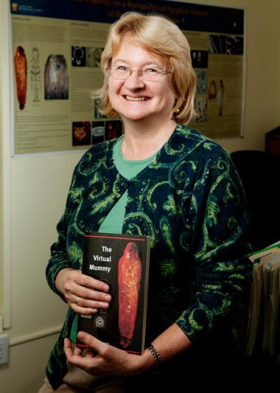
A team of medical experts and researchers present new findings on an ancient mummy in the collection of the University of Illinois Spurlock Museum. Sarah Wisseman, director of the Program on Ancient Technologies and Archaeological Materials at the Illinois State Archaeological Survey at Illinois and author of "The Virtual Mummy," led the effort to study the mummy without removing its shroud.
(Photo Credit: L. Brian Stauffer)
David Hunt, of the Smithsonian Institution's National Museum of Natural History, observed that the child still had some of its baby teeth, with adult teeth coming in. This and evidence that the long bones were still growing at the time of death indicate that the child was 7 to 9 years old, Wisseman said.
Several signs – including a cracked skull with no evidence of bleeding and the detection of carrion beetles in the body – suggest that the embalmers "did a crummy job or this body was lying around for a while before it was treated," Wisseman said. If the child died during an epidemic there could have been a lot of corpses to deal with, she said, causing delays or forcing the embalmers to rush.
"All of the evidence, however, suggests that this is a child from a wealthy family," she said. "They're using expensive red pigment from Spain. They're using gold gilt decoration. This is a fairly high-class kid."
Despite the high-tech probing, the mummy has maintained some of its secrets. Its hands are positioned in front its collapsed pelvis, hiding any evidence of its sex. And DNA tests of a sample collected from the damaged region near its base have yielded no definitive results so far.
There are some "tantalizing" clues to the child's sex in the face portrait attached to the mummy, Wisseman said, but such images can be misleading.
"There's a suggestion around the portrait of a tunic with a stripe on it. This alone would suggest that the child inside is a boy," she said. "But there are other mummies that have one person depicted on the outside and then you discover it's a different sex or even an animal instead of a human, so you can't tell a book by its cover."
The CT scans also revealed something that might be a lock of hair on one side of the child's head, Wisseman said.
"In the Roman period in Egypt, around A.D. 100, we do have examples of Roman face portraits with a shaved head and then a lock of hair on one side," she said. Boys had the lock on one side, girls on the other. But the evidence is not conclusive.
"We may not ever know whether the child was male or female," she said. "And we still don't know the cause of death."
The symposium, "The Return of the Mummy: New Imaging Results on the Spurlock Museum's Egyptian Mummy," will begin at 4 p.m. at the Knight Auditorium, Spurlock Museum, 600 S. Gregory St., Urbana. Admission is free.
Along with Wisseman, Barkmeier and Hunt, other members of the investigative team will speak at the symposium, including Dr. Allan Campbell, clinical professor of pathology and dermatology at the U. of I. College of Medicine at Peoria; Emily Teeter, a research associate at the Oriental Institute museum, University of Chicago; and Carter Lupton, curator of ancient history, Milwaukee Public Museum.
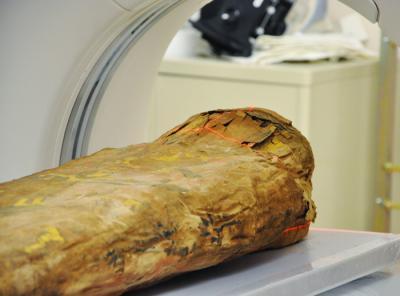
An analysis of CT scans and other studies indicate that the mummy was a child from a wealthy family in the Roman period of ancient Egypt.
(Photo Credit: Photo by Melissa Sotelo, Spurlock Museum)