One of the cytoskeleton components IRSp53 connects to is the tumor-promoting protein Eps8. IRSp53 is synergistically activated by the combined action of Cdc42 and binding of Eps8, which is upregulated in metastatic cancers.
Co-authors Tatyana Svitkina and Changsong Yang from the Penn Department of Biology, brought their expertise with living cells to the study. By introducing normal and mutant proteins into cells they could see how these proteins induced filopodia to form. The team found that mutations in critical regions of IRSp53 can either lead to enhanced or reduced filopodia formation and, as a consequence, cell motility. "This finding shows how all these different proteins converge on IRSp53 to execute precise cellular functions, and that when one factor is disrupted, other proteins are affected down the activity pathway," says Dominguez.
The team's next steps will be to screen libraries of small molecule inhibitors that interfere with the IRSp53-Eps8 interaction, to figure out how to stop unwanted cell movement before it gets too far.

Aberrant filopodia are induced by co-expression of fluorescently labeled Cdc42 and non-fluorescent IRSp53. Fluorescence shows the cell shape, because Cdc42 localizes to the plasma membrane.
(Photo Credit: Tatyana Svitkina, Ph.D., and Changsong Yang, Ph.D., Department of Biology, University of Pennsylvania)
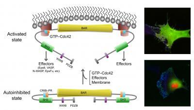
This image shows the active, open state of IRSp53 (top) and inactive, closed (bottom) state of IRSp53. In the closed state, cells do not generate filopodia as shown in the right bottom image (green=IRSp53, red=cdc42, and blue=actin, the most abundant protein in the cytoskeleton). The arrow between the two states indicates that the synergistic binding of Cdc42, cytoskeleton proteins (called downstream effectors of IRSp53 and includes the tumor-promoting factor Eps8) and the inherent attraction for the cell membrane, bring IRSp53 to specific locations on the cell membrane in which to change the shape of the cell. Chief among this reshaping activity is generating filopodia, the long thin objects coming off the cell in the top right image (color scheme as right bottom image). Note the change in pattern of the green and red show that IRSp53 and cdc42 are working together and moving to many different locations around the cell.
(Photo Credit: Roberto Dominguez, Ph.D., David Kast, Ph.D., Perelman School of Medicine, University of Pennsylvania)