MEDFORD/SOMERVILLE, Mass. (March 15, 2012) -- The genetic pathway that regulates the way cells align themselves relative to each other has been found to act as a "stop sign" that signals organisms when to halt cell growth, according to new research published by biologists at the Center for Regenerative and Developmental Biology in Tufts University's School of Arts and Sciences.
The research, available in Stem Cells and Development online in advance of final editing, sheds light on one of the primary challenges to developing new ways to induce regenerative repair: discovering how new tissue knows when to stop growing.
"The planar cell polarity (PCP) pathway is known to control alignment of cells within the organism, and errors in PCP result in a variety of syndromes," said Michael L. Levin, Ph.D., senior author on the paper and director of the Center. "We found that this pathway also acts as a signal that we can target to induce continued growth of nerve tissue during regeneration."
The researchers reported that in planarian flatworms, an important model of regeneration, inhibition of one or all of the Vangl2, DAAM1, and ROCK genes in the PCP pathway resulted in excess growth of the central nervous system, as well as extra eyes and excess optical neurons.
The Tufts biologists also found that PCP played a similar signaling role in vertebrates. Loss of Vangl2 led to overgrowth of neural tissue in Xenopus tadpoles during both normal development and tail regeneration.
Tissue growth is central to many biological processes, including embryonic development, routine cell replacement and regeneration following injury or disease. During tissue growth, stem and progenitor cells must be integrated and aligned with surrounding tissues and cells, and tissues must sense when that growth is complete. When such growth is not carefully controlled, the results can be disastrous.
"Right now very little is known about how new growth is halted during normal processes," said Levin. "Gaining control over cellular growth decisions is a crucial component of strategies designed to improve biological pattern formation and many related clinical advances. For example, in regenerative medicine we need to restore lost organs and limbs while maintaining both correct form and function."
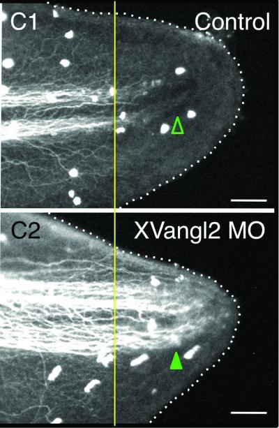
Immunofluorescent marking of Xenopus tadpole neurons shows that when the Vangl2 gene in the PCP pathway is interrupted, the developing tadpole exhibits 21 percent more nerve growth in the tail tip (bottom) compared with control (top). Open arrowheads indicate no nerve growth; solid arrowheads show increased nerve growth. Very little is known about how new cell growth is halted during normal processes. This discovery by Tufts University biologists has implications for areas such as tissue regeneration and wound healing, and prevention and treatment of cancer and birth defects.
(Photo Credit: Center for Regenerative and Developmental Biology at Tufts University)
"We hypothesized that the PCP pathway, which coordinates a variety of cellular behaviors in every living organism, could be involved in neuronal growth regulation on an organism-wide scale," said Wendy Scott Beane, Ph.D., first author on the paper and post-doctoral fellow in the Center.
Research by Beane and her colleagues on regeneration in planarians showed that loss of PCP through gene silencing resulted in continued neural growth for at least six weeks after the normal regeneration time of two weeks.
There was as much as a two-fold increase in excess neurons when an RNAi cocktail inhibiting the Vangl2, DAAM1, and ROCK genes was administered to regenerating flatworms. When single genes were targeted, 31.1% had excess eyes compared with 7.2% of controls. Extra pigment cells were also observed. PCP inhibition also caused excess neurons to form in intact planarians during normal cell replacement.
Examination using an immunofluorescent marker that recognizes planarian photoreceptor cells showed even greater overgrowth of optical nerves than observation of cell morphology alone: When single PCP genes were inhibited, 81% of the samples had excess optical neural tissue by eight weeks of regeneration; that increased to 100% when multiple genes were inhibited.
Furthermore, PCP inhibition in Xenopus tadpoles revealed PCP- mediated regulation of neuron growth during both regeneration and embryonic development. Together these results suggest that PCP signaling restricts neural growth during regeneration, homeostasis and development of vertebrates and invertebrates.
The researchers noted, however, that PCP inhibition did not change the flatworms' overall size or shape, suggesting that a master regulator of body shape still functioned.
In addition to Beane and Levin, co-authors of the paper included three other members of the Center on Regenerative and Developmental Biology: AiSun Tseng, Ph.D., postdoctoral fellow; Junji Morokuma, Ph.D., research associate, and Joan Lemire, Ph.D., research associate.
Source: Tufts University