Like tiny crawling compass needles, whole living cells and cell fragments orient and move in response to electric fields — but in opposite directions, scientists at the University of California, Davis, have found. Their results, published April 8 in the journal Current Biology, could ultimately lead to new ways to heal wounds and deliver stem cell therapies.
When cells crawl into wounded flesh to heal it, they follow an electric field. In healthy tissue there's a flux of charged particles between layers. Damage to tissue sets up a "short circuit," changing the flux direction and creating an electrical field that leads cells into the wound. But exactly how and why does this happen? That's unclear.
"We know that cells can respond to a weak electrical field, but we don't know how they sense it," said Min Zhao, professor of dermatology and ophthalmology and a researcher at UC Davis's stem cell center, the Institute for Regenerative Cures. "If we can understand the process better, we can make wound healing and tissue regeneration more effective."
The researchers worked with cells that form fish scales, called keratocytes. These fish cells are commonly used to study cell motion and they also readily shed cell fragments, wrapped in a cell membrane but lacking a nucleus, major organelles, DNA or much else in the way of other structures.
In a surprise discovery, whole cells and cell fragments moved in opposite directions in the same electric field, said Alex Mogilner, professor of mathematics and of neurobiology, physiology and behavior at UC Davis and co-senior author on the paper.
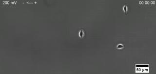
These fish cells crawl towards the negative electrode, or cathode, and change direction when the electric field is reversed. Electric fields may recruit or guide cells into wounded tissue.
(Photo Credit: Min Zhao and Alex Mogilner, UC Davis.)
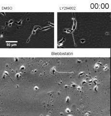
Both whole cells and cell fragments that break off move in an electric field, but in opposite directions. Electric fields may guide cells into wounded tissue to speed repair.
(Photo Credit: Min Zhao and Alex Mogilner, UC Davis.)
It's the first time that such basic cell fragments have been shown to orient and move in an electric field, Mogilner said. That allowed the researchers to discover that the cells and cell fragments are oriented by a "tug of war" between two competing processes.
Think of a cell as a blob of fluid and protein gel wrapped in a membrane. Cells crawl along surfaces by sliding and ratcheting protein fibers inside the cell past each other, advancing the leading edge of the cell while withdrawing the trailing edge.
Assistant project scientist Yaohui Sun found that when whole cells were exposed to an electric field, actin protein fibers collected and grew on the side of the cell facing the negative electrode (cathode) while a mix of contracting actin and myosin fibers formed toward the positive electrode (anode). Both actin alone, and actin with myosin, can create motors that drive the cell forward.
The polarizing effect set up a tug-of-war between the two mechanisms. In whole cells, the actin mechanism won and the cell crawled toward the cathode. But in cell fragments, the actin/myosin motor came out on top, got the rear of the cell oriented toward cathode and the cell fragment crawled in the opposite direction.
The results show that there are at least two distinct pathways through which cells respond to electric fields, Mogilner said. At least one of the pathways — leading to organized actin/myosin fibers — can work without a cell nucleus or any of the other organelles found in cells, beyond the cell membrane and proteins that make up the cytoskeleton.
Upstream of those two pathways is some kind of sensor that detects the electric field. In a separate paper to be published in the same journal issue, Mogilner and Stanford University researchers Greg Allen and Julie Theriot narrow down the possible mechanisms. The most likely explanation, they conclude, is that the electric field causes certain electrically charged proteins in the cell membrane to concentrate at the membrane edge, triggering a response.
The team also included Hao Do, Jing Gao and Ren Zhao, all at the Institute for Regenerative Cures and the UC Davis departments of Ophthalmology and Dermatology. Sun is co-advised by Mogilner and Zhao; Gao is now working at Yunnan Normal University, Kunming, China, and Ren Zhao is at the Third Military Medical University, Chongqing, China.
The work was funded by the National Institutes of Health, the California Institute for Regenerative Medicine and the National Science Foundation.