Using protein engineering and two different cutting-edge structural biology imaging techniques, researchers have developed a detailed picture of the protein largely responsible for enabling HIV to enter human immune cells and cause infection. An in-depth understanding of the atomic structure of the HIV envelope trimer—or Env, the three-component protein found on HIV's surface—is critical to better understanding how HIV gains entry into cells and for creating potential HIV vaccines.
Atomic-resolution imaging of the Env protein has previously been elusive because of the protein's complex, delicate structure. To capture the image, a team of scientists at The Scripps Research Institute and Weill Medical College of Cornell University engineered a more stable version of the protein. Then, in separate studies, using first cryo-electron microscopy and then X-ray crystallography, the researchers were able to reveal the structure of the Env trimer, how it assembles and how it interacts with broadly neutralizing antibodies that target HIV.
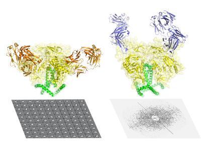
Shown here are cryo-electron miscroscopy (image 1) and X-ray crystallography (image 2) views of the HIV envelope protein.
(Photo Credit: The Scripps Research Institute)
Their research, described in two papers published online today in Science Express, received major support from the National Institute of Allergy and Infectious Diseases (NIAID), part of the National Institutes of Health.
Source: NIH/National Institute of Allergy and Infectious Diseases