Scientists at Johns Hopkins have turned their view of osteoarthritis (OA) inside out. Literally. Instead of seeing the painful degenerative disease as a problem primarily of the cartilage that cushions joints, they now have evidence that the bone underneath the cartilage is also a key player and exacerbates the damage. In a proof-of-concept experiment, they found that blocking the action of a critical bone regulation protein in mice halts progression of the disease.
The prevailing theory on the development of OA focuses on joint cartilage, suggesting that unstable mechanical pressure on the joints leads to more and more harm to the cartilage—and pain to the patient—until the only treatment option left is total knee or hip replacement. The new theory, reported May 19 in Nature Medicine, suggests that initial harm to the cartilage causes the bone underneath it to behave improperly by building surplus bone. The extra bone stretches the cartilage above and speeds its decline.
"If there is something wrong with the leg of your chair and you try to fix it by replacing the cushion, you haven't solved the problem," says Xu Cao, Ph.D., director of the Center for Musculoskeletal Research in the Department of Orthopaedic Surgery at the Johns Hopkins University School of Medicine. "We think that the problem in OA is not just the cartilage 'cushion,' but the bone underneath," he adds.
Joints are formed at the intersection of two bones. To prevent the grinding and wearing down of the ends of the bones, they are capped with a thin layer of cartilage, which not only provides a smooth surface for joint rotation but also absorbs some of the weight and mechanical strain placed on the joint. The degeneration of this protective layer causes extreme pain leading to limited mobility.
Cao says degeneration is most frequently initiated by instability in the load-bearing joints of the knee and hip caused by injury or strain, so athletes, overweight people and people whose muscles are weakened by aging are at highest risk of developing OA. The prevalence of the disease is rapidly increasing; it currently affects 27 million Americans and may double by 2030. The only treatment available is pain management, or surgical replacement of the arthritic joint with a prosthetic one.
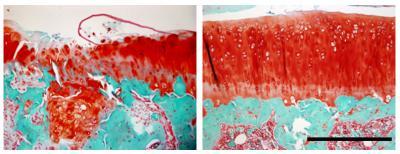
When placed in the bone (green) beneath the cartilage (red) of a rat's knee joint, antibodies against the protein TGF-beta1 can prevent the damage caused by osteoarthritis. Left, without treatment; right, with treatment.
(Photo Credit: Gehua Zhen, Courtesy of Nature Medicine)
Cao says that the lack of effective drugs or a complete understanding of the underlying process that causes OA to progress led his group to search for a different underlying cause. "We began to think of cartilage and the bone underneath it, called subchondral bone, as functioning as a single unit," says Cao. "That helped us to see the ways in which the bone was responding to changes in the cartilage and exacerbating the problem."
Using mice with ACL (anterior cruciate ligament) tears, which are known to lead to OA of the knee, the researchers found that, as soon as one week after the injury, pockets of subchondral bone had been "chewed" away by cells called osteoclasts. This process activated high levels in the bone of a protein called TGF-beta1, which, in turn, recruited stem cells to the site so that they could create new bone to fill the holes. Cao calls these pockets of new bone formation "osteoid islets."
But the bone building and the bone destruction processes were not coordinated in the mice, and the bone building prevailed, placing further strain on the cartilage cap. It is this extraneous bone formation that Cao and his colleagues believe to be at the heart of OA, as confirmed in a computer simulation of the human knee.
With this new hypothesis in hand, complete with a protein suspect, the team tried several methods to block the activity of TGF-beta1. When a TGF-beta1 inhibitor drug was given intravenously, the subchondral bone improved significantly, but the cartilage cap deteriorated further. However, when a different inhibitor of TGF-beta1, an antibody against it, was injected directly into the subchondral bone, the positive effects were seen in the bone without the negative effects on the cartilage. The same result was also seen when TGF-beta1 was genetically disrupted in the bone precursor cells alone.
"Our results are potentially really good news for patients with OA," says Cao. "We are already working to develop a clinical trial to test the efficacy of locally applied TGF-beta1 antibodies in human patients at early stages of OA." If successful, their nonsurgical treatment could make OA — and the pain and debilitation it causes — halt in its tracks, he says.
Source: Johns Hopkins Medicine