WASHINGTON, Sept. 13—By combining three previously unrelated imaging tools into one new device, a team of researchers from the University of Connecticut and the University of Southern California has proposed a new way to diagnose early-stage ovarian cancer in high-risk women through minimally invasive surgery. The new technique may be better than the current standard procedure of preemptively removing the ovaries.
Ovarian cancer has a low survival rate because a lack of reliable screening techniques usually means the disease remains hidden until the later stages. Now researchers have drawn on the unique advantages of multiple imaging tools to test a new way of spotting early-on the tissue irregularities that signal cancer.
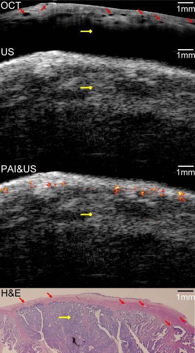
Co-registered images of malignant ovarian tissue obtained with the hybrid imaging device. From top to bottom: OCT image , ultrasound image, superimposed photoacoustic and ultrasound image and corresponding histology . Yellow diamond arrow: malignant tissue.
(Photo Credit: University of Connecticut/Biomedical Optics Express)
For their diagnostic device, the researchers combined the contrast provided by photoacoustic imaging, the high-resolution subsurface imaging provided by optical coherence tomography, and the deeper tissue imaging provided by pulse-echo ultrasound. They tested their device, described by the team in the September issue of the Optical Society's (OSA) open-access journal Biomedical Optics Express, by imaging both pig and human ovarian tissue, and correctly identified malignant tumors that were later confirmed by staining the tissue and examining it under a microscope. These initial tests were performed on tissue that had been surgically removed, but the diameter of the device – at only 5 mm – is small enough that it could potentially be inserted through a small slit to image tissue in live patients.
Source: Optical Society of America