OAK BROOK, Ill. – Researchers using a combination of different imaging techniques have found structural abnormalities in the brains of people with chronic fatigue syndrome (CFS), according to a new study published online in the journal Radiology. The results suggest a potential role for imaging in diagnosing and treating the condition.
CFS is characterized by profound fatigue and "brain fog" that do not improve with bed rest, lasting for at least six months. The condition affects more than 1 million adults and children in the United States, according to the Centers for Disease Control and Prevention. Diagnosis is complicated and usually involves ruling out many other conditions. There is no standalone test to diagnose CFS.
"This is a very common and debilitating disease," said the study lead author Michael M. Zeineh, M.D., Ph.D., assistant professor of radiology at Stanford University School of Medicine in Stanford, Calif. "It's very frustrating for patients, because they feel tired and are experiencing difficulty thinking, and the science has yet to determine what has gone wrong."
For the new study, Dr. Zeineh worked with a Stanford CFS and infectious disease expert, Jose G. Montoya, M.D., to perform magnetic resonance imaging (MRI) on 15 CFS patients and 14 age- and gender-matched controls. They applied three different MRI techniques: volumetric analysis to measure the size of different compartments of the brain, diffusion tensor imaging (DTI) to assess the integrity of the signal-carrying white matter tracts of the brain, and arterial spin labeling (ASL) to measure blood flow.
When they compared results between the CFS patients and the controls, they found that the CFS group had slightly lower white matter volume, meaning there was less overall white matter in the brain. The CFS group also had abnormally high fractional anisotropy (FA) values—a DTI measure of the diffusion of water— in a specific white matter tract called the right arcuate fasciculus, suggesting something was going on in the white matter in the right hemisphere.
"Within CFS patients, right anterior arcuate FA increased with disease severity," Dr. Zeineh said. "The differences correlated with their fatigue—the more abnormal the tract, the worse the fatigue."
The results suggest that FA at the right arcuate fasciculus may serve as a biomarker for CFS that can help track the disease.
The imaging study also found abnormalities among CFS patients at the two points in the brain that connect the right arcuate fasciculus. Each connection point, known as a cortex, was thicker in CFS patients.
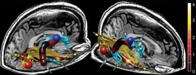
This reconstructed MR image shows the right arcuate (blue tracks and arrows) and ILFs (yellow tracks and arrows) in a single representative subject. These two tracks are overlaid on their respective track profiles. The track profile is colored according to the T score of track-based FA, showing that the maximal increase in FA is in the anterior arcuate and ILFs. The red, blue, and green spheres correspond to size and locations of increased cortical thickness from Figure 1 in the right occipital, precentral, and middle temporal regions, respectively. The green arrows also point to the middle temporal region of increased thickness.
(Photo Credit: Radiological Society of North America)
"This is the first study to look at white matter tracts in CFS and correlate them with cortical findings," Dr. Zeineh said. "It's not something you could see with conventional imaging."
Although the study involved only 15 CFS patients, the technique already shows tremendous promise as a diagnostic tool for identifying people with CFS, according to Dr. Zeineh.
"We used automated techniques to look at these tracts and were able to achieve 80 percent accuracy for CFS detection," he said.
Dr. Zeineh added that the findings need to be replicated and expanded upon in future studies to refine understanding of the relationship between brain structure and CFS.
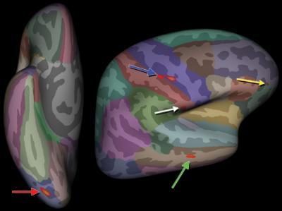
Superimposed on the inflated atlas brain image are regions of right-hemisphere increased cortical thickness in patients with CFS compared with control subjects after accounting for differences in age and handedness are highlighted in red (arrows). Blue = precentral, green = middle temporal, red = occipital, white = postcentral, and yellow = orbitofrontal. The right precentral region consists of two con¬nected foci that FreeSurfer identified as one contiguous region.
(Photo Credit: Radiological Society of North America)