At CLEO: 2014, being held June 8-13 in San Jose, California, USA, UTHealth scientist John Rasmussen will describe how they have taken this technology, which they call near-infrared fluorescence lymphatic imaging (NIRFLI), from bench-top development to various clinical applications.
"We feel that the ability to see the lymphatics will provide opportunities to revolutionize lymphatic care," Rasmussen said.
Why the Human Lymphatic System is Hard to Image
The major problem with lymphatic imaging is that the small lymphatic vessels are filled with lymph, a clear liquid that lacks the natural contrast needed to show up on instruments like CT scanners or MRIs. While one might think about injecting dyes or other contrast "agents" into the lymphatic vessels to make them more visible, the vessels are very difficult to find and are most often too small to insert a needle.
An existing technology, called lymphoscintigraphy, can take images of the lymphatic system following injection of a radioactive compound into or below the skin. However, lymphoscintigraphy typically takes 20-45 minutes to acquire a single grainy picture, and can only image the largest lymphatic vessels or trunks. The smaller vessels, which make up the bulk of the lymphatic system, are invisible to lymphoscintigraphy. In addition because of the long acquisition times, it cannot capture the real-time flow of fluid in the system.
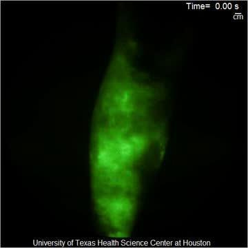
This is a movie illustrating the lack of lymphatic flow in the lower leg of a subject with lymphedema.
(Photo Credit: John Rasmussen)
At CLEO: 2014, Rasmussen will describe how near-infrared fluorescence laser imaging (NIRFLI) allows lymphatic structures and flow to be measured quantitatively, including in the fine vessels—a dramatic improvement over lymphoscintigraphy. In addition, NIRFLI will be safer and less expensive than existing technology.
To acquire images of the lymphatics, NIRFLI uses indocyanine green dye, which is injected in tiny amounts into the skin of a patient. The dye is absorbed into the lymphatics and when illuminated by the laser diode, it emits a fluorescent light, which the device amplifies with a military-grade image intensifier—the main component in night vision goggles—and then captures with a commercial CCD digital camera.
The image intensifier enables the small lymphatic vessels to be visualized, and by taking sequences of such images, they can produce movies showing flow within the lymphatics. Rasmussen said that the most immediate promise of NIRFLI will be to diagnose and monitor the treatment of lymphedema and may also help surgeons identify and remove lymph nodes into which cancer tumors drain.
"From these images and movies, we can identify abnormal lymphatic structure and function in a variety of diseases and disorders in which the lymphatics play a role," Rasmussen said. "I think we have barely scratched the surface of what is possible."
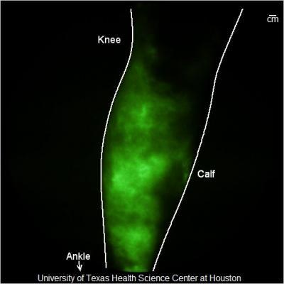
This is a typical near-infrared fluorescence image of lymphatics in the lower leg of a subject with lymphedema.
(Photo Credit: John Rasmussen)
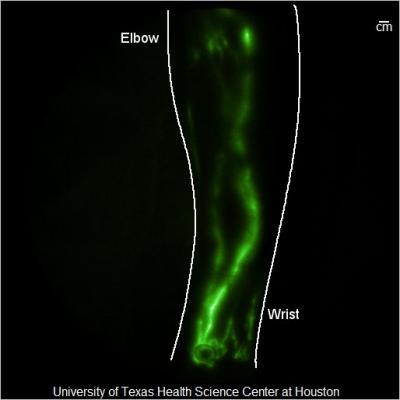
This is a typical near-infrared fluorescence image of healthy lymphatics in the lower arm.
(Photo Credit: John Rasmussen)
Source: The Optical Society