Currently the resolution of the images is not as fine as what can be obtained with existing breast imaging techniques like X-ray mammography and MRI. In future versions, Srirang Manohar, an assistant professor at the University of Twente who led the research, Wenfeng Xia, a graduate student at the University of Twente who is the first author on the new paper, and their colleagues expect to improve the resolution as well as add the capability to image using several different wavelengths of light at once, which is expected to improve detectability.
The Twente researchers, who belong to the Biomedical Photonic Imaging group run by Professor Wiendelt Steenbergen, have tested their prototype in the laboratory using phantoms -- objects made of gels and other materials that mimic human tissue. Last year, in a small clinical trial they showed that an earlier version of the technology could successfully image breast cancer in women.
Manohar and his colleagues added that if the instrument were commercialized, it would likely cost less than MRI and X-ray mammography.
"We feel that the cost could be brought down to be not much more expensive than an ultrasound machine when it goes to industry," said Xia.
The next step, they say, will be to prepare for larger clinical trials. Several existing technologies are already widely used for breast cancer screening and diagnosis, including mammography, MRI, and ultrasound. Before becoming routinely used, the photoacoustic mammoscope would have to prove at least as effective as those other techniques in large, multicenter clinical trials.
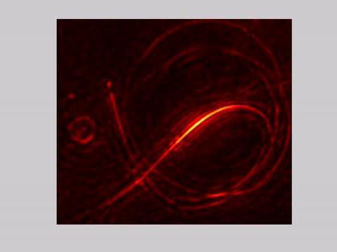
This video shows the rotation of the reconstructed phantom as seen from above.
(Photo Credit: Wenfeng Xia, Biomedical Photonic Imaging group, University of Twente)
"We are developing a clinical prototype that improves various aspects of the current version of the device," said Manohar. "The final prototype will be ready for first clinical testing next year."
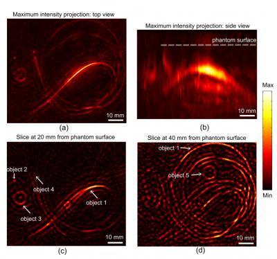
The top image shows a top and side view of a 3-D-reconstructed phantom object made of gels and other materials that mimic human tissue. The background of this phantom mimics normal breast tissue, while several objects embedded within the material mimic blood vessels and tumors.The bottom image shows two slices of images of a reconstructed phantom taken with the new device. The location of five objects are indicated with arrows: objects 1 and 2 mimic blood vessels, while objects 3-5 mimic tumors.
(Photo Credit: Wenfeng Xia, Biomedical Photonic Imaging group, University of Twente)
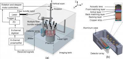
This is a schematic illustration of the imaging system and the ultrasound detector.
(Photo Credit: Wenfeng Xia, Biomedical Photonic Imaging group, University of Twente)
Source: The Optical Society