The acetylcholine system plays a pivotal role in working memory, holding information in mind temporarily, but appears to act by influencing the processing of information rather than through memory. Imaging studies suggest that visual working memory performance can be enhanced by modulating acetylcholine-induced activity in the brain's visual processing area, called the visual cortex, when processing information that is important to the task. Since working memory performance can predict response to conventional antidepressants and ketamine, Furey and colleagues turned to a working memory task and imaging visual cortex activity as potential tools to identify a biomarker for scopolamine response.
Depressed patients have a well-known tendency to process and remember negative emotional information. The researchers propose that this bias stems from dysregulated acetylcholine systems in some patients. They reasoned that such patients would show aberrant visual cortex activity in response to negative emotional features of a working memory task. They also expected to find that patients with more dysfunctional acetylcholine systems would respond better to scopolamine treatment.
Before receiving scopolamine, participants performed a working memory task while their brain activity was monitored via fMRI. For some trials, it required that they pay attention to, and remember, the emotional expression (sad, happy, etc.) of faces flashing on a computer monitor. For other trials, they had to pay attention to only the identity, or non-emotional feature, of the faces. After scanning, and over the following several weeks, 15 patients with depression and 21 healthy participants randomly received infusions of a placebo (salt solution) and/or scopolamine. Mood changes were monitored with depression rating scales.

NIMH's Dr. Maura Furey talks about how a functional brain imaging measure may help predict a patient's response to a rapid-acting experimental antidepressant.
(Photo Credit: National Institute of Mental Health)
Overall, scopolamine treatment reduced depression symptoms by 63 percent, with 11 of the patients showing a significant clinical response. The strength of this response correlated significantly with visual cortex activity during key phases of the working memory task – while participants were paying attention to the emotional content of the faces. There was no such correlation for trials when they attended to the identity of the faces.
The findings suggest that acetylcholine system activity drives visual cortex activity that predicts treatment response – and that differences seen between depressed patients and controls may be traceable to acetylcholine dysfunction. Overall, patients showed lower visual cortex activity than controls during the emotion phase of the task. Patients showing activity levels most dissimilar to controls experienced the greatest antidepressant response to scopolamine treatment. Visual cortex activity in patients who didn't respond to scopolamine more closely resembled that of controls. As hypothesized, the pretreatment level of visual cortex activity appears to reflect the extent of patients' acetylcholine system dysfunction and to predict their response to the experimental medication, say the researchers.
Preliminary evidence suggests that such visual cortex activity in response to emotional stimuli may also apply to other treatments and may prove to be a shared biomarker of rapid antidepressant response, according to Furey.
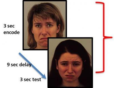
Over several trials, participants were required to attend to either the identity (non-emotional feature) or the emotion of a face, remember it during a 9-second delay, and match the feature to a subsequent face. Neural activity in the visual cortex elicited by the emotion trials predicted depressed patients' subsequent responsiveness to scopolamine treatment.
(Photo Credit: Maura Furey, Ph.D., NIMH Experimental Therapeutics and Pathophysiology Branch)
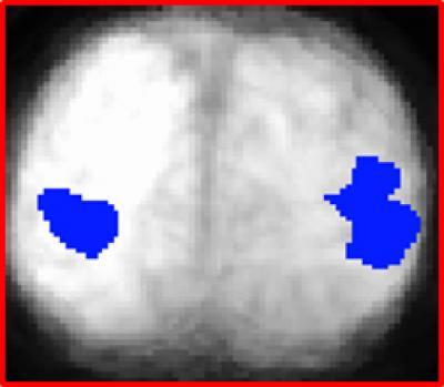
The level of increased activity in left and right visual cortex (blue), while attending to emotional faces in a working memory task, predicted depressed patients' responsiveness to the experimental antidepressant scopolamine. View from the back of the brain shows fMRI data superimposed on anatomical MRI scan data.
(Photo Credit: Maura Furey, Ph.D., NIMH Experimental Therapeutics and Pathophysiology Branch)