Going forward, the researchers would like to increase the field of view of the device so that they can image the full lesion along with its border to healthy tissue. They are also working on speeding up the post-processing of the optical signal to enable live vasculature display, and improving the portability of the system, which currently occupies an area about half the size of an office desk. "We believe that in the future our method will help to simplify non-invasive dermatological in vivo diagnostics and allow for in-depth treatment monitoring," says Blatter.
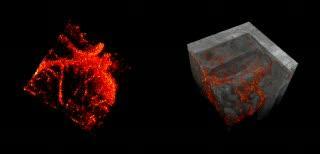
This is a 2x2x2mm extracted blood vessel structure of basal cell carcinoma in vivo exhibiting a chaotic vascular pattern (left). Virtually cut volume obtained with OCT displaying the embedded blood vessels in red (right).
(Photo Credit: Courtesy Medical University Vienna)
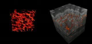
This is a 2x2x2mm extracted blood vessel structure in 3-D of healthy skin in vivo obtained with the label-free optical motion contrast method (left). Virtually cut volume obtained with OCT displaying the embedded blood vessels in red (right).
(Photo Credit: Optics Express (2011))
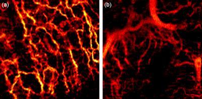
Image (a) on the left shows a healthy network of blood vessels in the lower layer of skin on the hand palm. Image (b) shows blood vessels supplying a basal cell carcinoma on the forehead. The effects of disease on the vascular are evidenced by the branching pattern of vessels in image (b), with abnormally large vessels for a depth range similar to image (a). Both images show a 2x2 millimeter area.
(Photo Credit: Medical University Vienna/ Biomedical Optics Express)
Source: Optical Society of America