Researchers don't even really know if invadopodia have a normal purpose in healthy tissue. Pack explains that their study was the first to show that invadopodia occur in living animals. This work in live zebrafish is the first direct evidence that invadopodia play a role in tissue cell invasion and identifies the physical signal that drives this process, which in turn, does so by inducing oxidative stress in the invasive cells.
Indeed, eliminating Tks5, a protein required for invadopodia formation in mammalian cells that helps generate reactive oxygen species, blocked formation of the protrusions and rescued mutant zebrafish from cell invasion.
Mouse studies along the same research lines as the zebrafish are planned. The zebrafish and mouse models under development could one day be used to screen potential drugs to stop formation of the errant invadopodia, thus preventing metastases at an early stage of their development. Supporting this idea, mutations in the human smooth muscle myosin genes are present in patients with colorectal cancer and are associated with cancer invasion.
In addition, the team's experiments also point to the possibility that humans who carry a single copy of smooth muscle mutations similar to the zebrafish mutation might inherit a disposition to cancer progression, if they develop a cancer independently of the mutation. The fish with one copy of the mutant gene develop normally but show cell invasion when exposed to drugs that cause oxidative stress, and the risk is greater in fish carrying oncogenes. "This is an interesting finding because oxidative stress is common in cancers and inflammatory conditions that predispose to cancer, such as ulcerative colitis," notes Pack.

The movie shows a segment of the mutant intestine (3.5 day old fish, lateral view of the intestine). The time lapse images were taken over about six to eight hours. It is a transgenic mutant zebrafish that expressed the green fluorescent protein tagged to a peptide that binds to actin in epithelial cells. Normally actin lines the portion of the intestinal cells that are adjacent to the lumen of the intestinal tube. This is the bright green staining in the intestine. In the mutants, there also are small actin rich protrusions that appear within the plasma cell membrane on opposite side of the cells undergoing invasion. Invasive cells with these protrusions expand out in to the surrounding tissue stroma (arrows). The green structure on the top of the movie frame is the kidney duct. The intestine is below the kidney duct. The actin-rich protrusions are the invadopodia. Transcriptome profiles of the cells with invadopodia showed that genes responsive to reactive oxygen species were upregulated in young mutant fish.
(Photo Credit: Christoph Seiler and Michael Pack, Perelman School of Medicine, University of Pennsylvania)
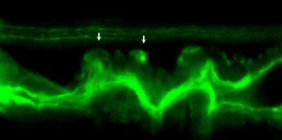
This is a segment of the mutant intestine (3.5 day old fish, lateral view of the intestine) in a transgenic mutant zebrafish that expressed the green fluorescent protein tagged to a peptide that binds to actin in epithelial cells. Invasive cells with protrusions expand out in to the surrounding tissue stroma (arrows). The green structure on the top of the movie frame is the kidney duct. The intestine is below the kidney duct. The actin-rich protrusions are the invadopodia.
(Photo Credit: Christoph Seiler and Michael Pack, Perelman School of Medicine, University of Pennsylvania)