Diffusion-tensor imaging can be used to observe the microstructure of brain tissue. Fractional anisotropy reflects the integrity of white matter fibers. Fractional anisotropy of a young adult brain is low in gray matter, high in white matter, and highest in the splenium of the corpus callosum. A recent study by Dr. Jun Pu and colleagues from Second Affiliated Hospital of Kunming Medical University in China investigated anisotropy values in major white matter tracts and gray matter nuclei in the brains of healthy adult rhesus monkeys. Results showed no laterality differences in fractional anisotropy values. Fractional anisotropy values were low in the head of caudate nucleus and thalamus in gray matter. Fractional anisotropy values were highest in the splenium of corpus callosum in the white matter, followed by genu of the corpus callosum and the posterior limb of the internal capsule. Fractional anisotropy values were lowest in the semioval center and posterior limb of internal capsule. These results suggest that fractional anisotropy values in major white matter fibers and the deep gray matter of adult rhesus monkeys are similar to those of healthy young people. These findings were published in the Neural Regeneration Research (Vol. 8, No. 31, 2013).
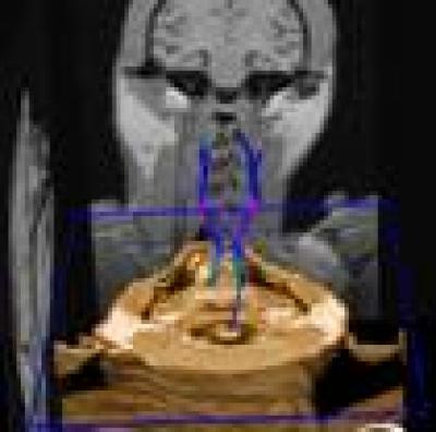
This corticospinal tract from a monkey brain can be seen to originate in the precentral gyrus, continue down through the internal capsule, and then down to the midbrain and medulla oblongata. Each fiber tract is continuous and uniform in the shape.
(Photo Credit: Neural Regeneration Research)
Source: Neural Regeneration Research