A team of scientists from The University of Texas at Arlington and MIT has figured out how to quantitatively observe cellular processes taking place on so-called "lab on a chip" devices in a silicon environment.
The new technology will be useful in drug development as well as disease diagnosis, researchers say.
In a paper published in Nature's online journal Scientific Reports, the team said it overcame past limitations on quantitative microscopy through an opaque media by working with a new combination of near infrared light and a technique called quantitative phase imaging. Quantitative phase imaging is about a decade old. It uses shifts in phases of light, not staining techniques, to aid specimen imaging – earning the term "label-free."
"To the best of our knowledge, this is the first demonstration of quantitative phase imaging of cellular structure and function in silicon environment," said Assistant Professor of Physics Samarendra Mohanty, head of the Biophysics and Physiology Laboratory at UT Arlington and corresponding author on the paper.
The UT Arlington/MIT team was able to prove success in analyzing specimens through a silicon wafer in two instances. In one, they accomplished full-field imaging of the features of red blood cells to nanometer thickness accuracy. In another, they observed dynamic variation of human embryonic kidney cells in response to change in salt concentration. Mohanty believes that his group's current work on near infrared quantitative phase imaging can lead to non-invasive, label-free monitoring of neuronal activities.
Additional co-authors include: Bipin Joshi and Nelson Cardenas, of UT Arlington; and Ishan Barman, Narahara Chari Dingari, Jaqueline S. Soares and Ramachandra R. Dasari, all of MIT.
"Silicon-based micro devices known as labs-on-a-chip are revolutionizing high throughput analysis of cells and molecules for disease diagnosis and screening of drug effects. However, very little progress has been made in the optical characterization of samples in these systems," said Joshi, a recent graduate and lead author on the paper. "The technology we've developed is well-suited to meet this need."
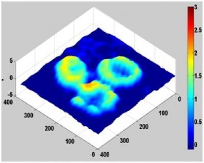
A UT Arlington/MIT team was able to prove success in analyzing specimens through a silicon wafer in two instances.
(Photo Credit: MIT)
Barman, now an assistant professor at Johns Hopkins University, said the new paper is a prime example of the type of research he hopes to do - projects pulled by needs of the biomedical community and continually pushing the edge of biophotonic solutions.
"We envision that this significantly expands the visualization possible in silicon based microelectronic and micromechanical devices," he said.
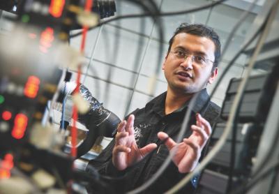
Assistant Professor of Physics Samarendra Mohanty, head of the Biophysics and Physiology Laboratory at UT Arlington, was corresponding author on a new paper in Scientific Reports.
(Photo Credit: UT Arlington)
Source: University of Texas at Arlington