"It's like Photoshop for cell biologists," Cohen said. "The software outlines cells and blood vessels, keeping track of them as they're dividing and moving around one another. This provides a wealth of information on the patterns of cell shape, motion and division. Visualization of the 3-D microscopy data together with the analysis results is a key step to measure and ultimately understand what drives these cells."
With an enhanced version of the program called LEVER 3-D, which compiles data from multi-layered microscopic images, Cohen's team can produce a three-dimensional rendering of the cells and animate it through time to show their multiplication and movement. The enhanced imaging gives researchers a unique view of the interaction of stem cells with their surroundings.
By running the software on a computer with a graphics card optimized for gaming and donning a set of 3-D gaming glasses, the program can take scientists inside the microscopic cross section. In a portion of Cohen's lab modified to be a viewing theater, a special stereo-vision projector brings the three-dimensional image data to life. Users can rotate and zoom in and out of the projected image in true 3-D, giving scientists vantage points that are not possible when looking through a microscope.
"LEVER 3-D is amazing, it opens new vistas for understanding the stem cell niche," said Dr. Sally Temple, a cell biologist at the Neural Stem Cell Institute in Rensselaer, N.Y. who has been using Cohen's software, through the course of its development, as part of her stem cell research since 2005. Cohen's goal is to make the software open-source and readily available to any scientists who can use it for their research.
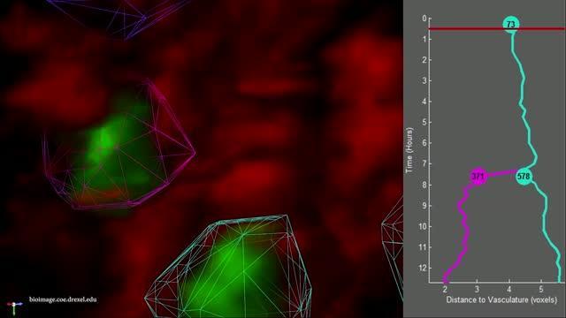
A software program developed by engineers at Drexel University, called Lineage Editing and Validation (LEVER), allows biologists to tag and track cell proliferation to validate lineage trees.
(Photo Credit: Drexel University)
The project, which is funded by the National Institute on Aging, was recently presented by Eric Wait, a doctoral researcher in Cohen's lab, at the Symposium on Biological Data Visualization in Boston. The team will continue to add interactive capabilities to their system as it becomes more widely used as a research tool.
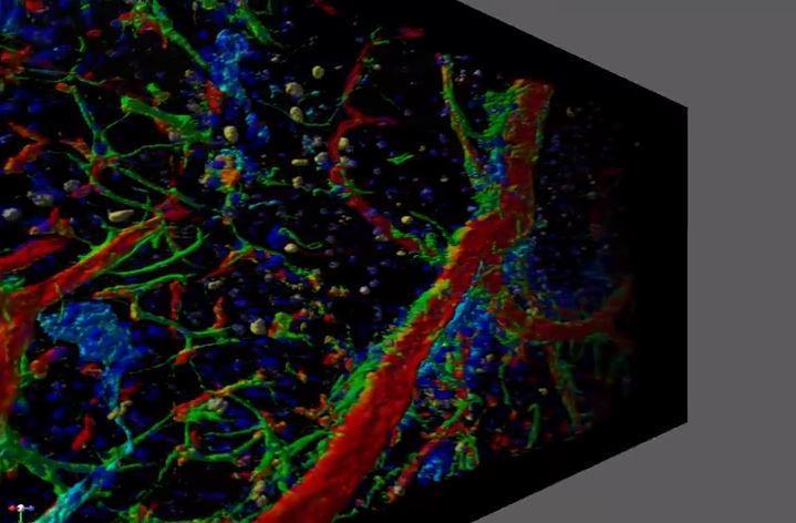
Drexel engineers created a program that can stack slices of microscopic time-lapse video in a three-dimensional volume. The one shown here is a cross section of brain tissue that shows stem cells (purple) and surrounding vasculature (red and green).
(Photo Credit: Drexel University)
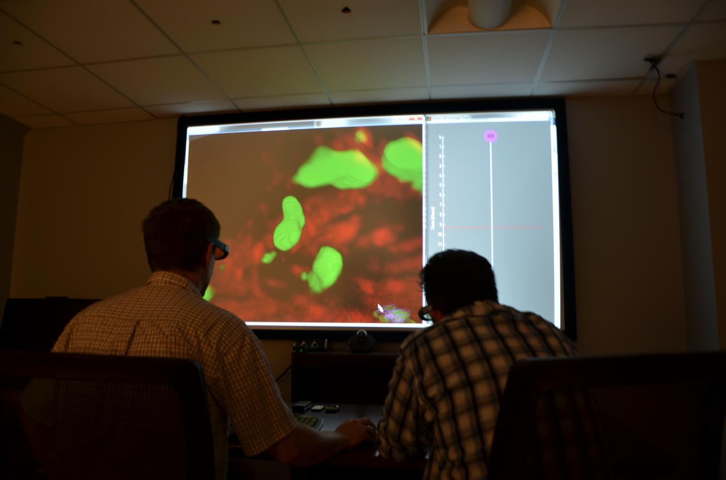
Using commercial home theater equipment and 3D stereoscopic gaming glasses, Drexel engineers turned their lab into a 3D movie theater for studying cells.
(Photo Credit: Drexel University)
Source: Drexel University