Infection with Coccidioides can progress through three stages of increasing severity. Valley fever is the acute form of the disease, which, if left untreated, can develop into a second-stage chronic infection, lasting months or years. This form affects roughly 40 percent of those exposed. The third stage of the disease, known as disseminated Coccidioides, occurs when the infection spreads throughout the body, affecting skin, bones and nervous system and causing skin ulcers, swollen joints and severe pain, abscesses, bone lesions, heart inflammation, urinary tract infection and (potentially lethal) meningitis. Disseminated Coccidioides affects 5-10 percent of those with chronic infection.
The rapid rise in valley fever cases in the arid southwest has become a serious health concern, as human habitation has pushed further into desert areas where the soil spores are widespread. Currently, Valley Fever affects an estimated 150,000 people a year, with most cases occurring in Arizona, California, Nevada, New Mexico and Utah. The disease has no cure at present and is notoriously tricky to diagnose. One reason is that valley fever is readily confused with other community-acquired pneumonias.
Currently, diagnosis is carried out through a technique known as immunodiffusion, which tests the blood for antibodies against Coccidioidal antigens. As the authors note, such tests are less than satisfactory, with a false negative rate as high as 50-70 percent. Around 5 percent of symptomatic patients display no measurable antibody levels to Valley Fever by immunodiffusion.
The current study describes an alternate method used to address the poor accuracy of immunodiffusion, applying an innovative new technique known as 'Immunosignaturing'. The technique can produce a detailed profile of system-wide immune activity from a small droplet of blood—typically, less than a microliter.
To produce its detailed immune portrait or immunosignature, the technique uses a microarray platform. This consists of a glass slide imprinted with 10,000 peptides. Each peptide consists of a string of 20 amino acids, randomly arranged. The power of the technology resides in the fact that the randomly generated peptides are not based on natural antigens to Coccidioides or indeed, any disease. They are "unbiased" to the nature of particular disease antibodies and can therefore act as a sort of universal diagnostic.
When a droplet of antibody-containing blood is smeared across the microarray, the random peptides behave like naturally occurring antigens, binding with blood antibodies in a specific pattern. Global analysis of the resulting immunosignature is used to establish disease-specific blueprints of immune activity.
The method potentially offers much higher resolution and sensitivity to disease, compared with diagnostic tests measuring a single antibody-antigen binding event or a small ensemble of molecules.
In the first round of experiments in the current study, the group used immunosignatures to determine if Valley Fever infected individuals could be accurately distinguished from three other patient groups afflicted with bacterial or fungal infections.
Once an immunosignature for Valley Fever was established using the 10K peptide microarray, a smaller diagnostic array was composed from relevant diagnostic peptides. This smaller 96-peptide array was then tested for accuracy against the current immunodiffusion diagnostic standard.
The 10K peptide array successfully distinguished Valley Fever from 3 other infections, with 98 percent accuracy. Impressively, the method also was able to classify false negative Valley Fever patients in a blinded test, with 100 percent accuracy, easily outpacing existing immunodiffusion methods, which could only identify 28 percent of false negatives.
The smaller, 96 peptide diagnostic array showed less specificity than the 10K peptide array in terms of identifying false negatives. The authors propose that the larger 10K peptide array be used in initial screenings, followed by subarrays with reduced complements of carefully selected peptides, used for clinical diagnosis.

In this video, biodesign researcher Krupa Navalkar describes a new diagnostic technique for pinpointing Valley Fever.
(Photo Credit: The Biodesign Institute at Arizona State University)
Immunosignaturing holds of promise for rapid, cost-effective and highly accurate diagnosis of Valley Fever. The versatile platform has the potential to separate Valley Fever patients from those afflicted with other bacterial or fungal infections. Making use of the same microarray, researchers can also identify false negatives with 100 percent accuracy.

Krupa Navalkar is a researcher at the Biodesign Institute at Arizona State University.
(Photo Credit: The Biodesign Institute at Arizona State University)
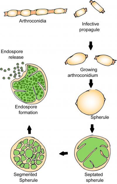
This graphic outlines the life cycle of Coccidioides, a fungal pathogen responsible for Valley Fever.
(Photo Credit: Public Domain)
Source: Arizona State University